With industry-leading sterile filtration devices based on 60 years of membrane filtration expertise, MilliporeSigma supports conventional and emerging formats for cell culture. The MilliporeSigma Cell Culture Solutions portfolio features over 20,000 high-quality tools to cultivate consistency in your cell culture workflow.
Cell Preparation: Tools that you need to minimize contamination risk and maximize workflow efficiency while preparing your media for cell culture.
Cell Growth: Whatever your cell culture growth needs, we have a plate, insert or vessel that can help you obtain the optimal cell culture results.
Cell Analysis: In addition to our solid base in assay tools and products, we offer new, novel cell culture technologies and cell culture platforms for high content data collection and analysis.
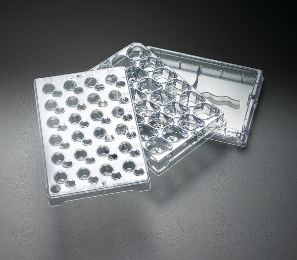
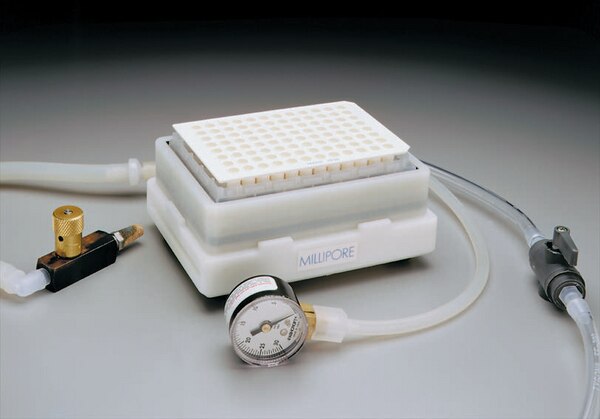
Millicell cell culture insert plates are a patented device designed to support cell growth and differentiation of endothelial and epithelial cell lines, including CaCo-2. The Millicell device is available with a 0.4 µm PCF and a variety of PET membrane pore sizes. The plate is designed for maximum user convenience and incorporates features such as apical assist for easier pipetting and basolateral access. To reduce the risk of monolayer contamination, the membrane plates have feet to elevate the plate above the work surface when disassembled from the feeder tray. Tear drop-shaped receiver wells eliminate air bubbles as plates are assembled.Millicell plate are optimized to grow and sustain high integrity cell monolayers. Cells grown on Millicell plate membranes grow better than on plastic because the cells are nourished from both the apical and basolateral sides. Cell growth and function more closely mimic in vivo. Millicell plates are designed for analysis as well as cell growth and can be used manually or with automated cell seeding, feeding and washing systemsWith this complete system, you can grow, feed, and analyze cells in one membrane-bottom plate. The plate can be used with either a single-well or a multi-well feeding tray. At the time of transport analysis, the plate is simply transferred to a multi-well transport tray for analysis. This streamlined design enhances functionality with: - Seed-and-feed systems - Automated liquid handling systems and span pipettes - Basolateral and apical access to cells - Transepithelial electrical resistance (TEER) measurement systems
Catalogue Number
PSHT010R5
Chemistry
Polycarbonate (PC)
Components
24-well cell culture plate, single-well feeder tray, lid
With Millicell inserts, attachment or suspension cells can access media from both their apical and basolateral sides. Cell growth, structure, and function more closely mimic what occurs in vivo. In addition, Millicell inserts make it possible to study both sides of the cell monolayer.Millicell inserts are available for 24-, 12-, or 6-well plates. The inserts are easily prepared for SEM and TEM visualizing techniques, and they are compatible with cellular and/or fluorescent stains.Insert Formats:Millicell Hanging Inserts: Preloaded in receivers or available standalone - For co-culturing and permeability assays - Unique design allows easier basolateral access than other hanging inserts with less risk of contamination - Available in 5 pore sizes and 3 diameters, including a 1 µm pore size that is optically transparent for better visualization by microscopy - NEW: Available preloaded in 24-well receiver plates or as individual inserts in 3 diameters Millicell Standing Inserts: - Promotes excellent cell growth and provides an exceptional opportunity for cell studies - Available with Biopore (PTFE) membrane, MF-Millipore (mixed cellulose esters) membrane, and polycarbonate membrane Millicell Organotypic Insert: - For high cell viability and superior study of three dimensional explant structure - Lower height allows them to fit inside a standard petri dish - The Biopore™ (PTFE) membrane provides high viability—for as long as 40 days—and excellent trans-membrane oxygen transport - The membrane is optically clear and optimized for long-term organotypic explant maintenance Membrane Types:Biopore Membrane (hydrophilic PTFE) - For low protein-binding, live cell viewing, and immunofluorescent applicationsMF-Millipore™ Membrane (mixed cellulose esters) - For exceptional anatomical and functional polarizationIsopore™ Membrane (polycarbonate) - For growth of attachment-dependent cells without matrixPET Membrane (polyethylene terephthalate) - For growth of attachment-dependent cells without matrixWith Millicell® inserts, adherent or suspension cells can access media from both their apical and basolateral sides. Cell growth, structure, and function more closely mimic what occurs in vivo. In addition, Millicell® inserts make it possible to study both sides of the cell monolayer.ApplicationsCell Attachment, Cell Growth, Cell Differentiation , Immunocytochemistry
Catalogue Number
PIHP01250
Chemistry
References
Product Information
Device Configuration
With Millicell inserts, attachment or suspension cells can access media from both their apical and basolateral sides. Cell growth, structure, and function more closely mimic what occurs in vivo. In addition, Millicell inserts make it possible to study both sides of the cell monolayer.Millicell inserts are available for 24-, 12-, or 6-well plates. The inserts are easily prepared for SEM and TEM visualizing techniques, and they are compatible with cellular and/or fluorescent stains.Insert Formats:Millicell Hanging Inserts: Preloaded in receivers or available standalone - For co-culturing and permeability assays - Unique design allows easier basolateral access than other hanging inserts with less risk of contamination - Available in 5 pore sizes and 3 diameters, including a 1 µm pore size that is optically transparent for better visualization by microscopy - NEW: Available preloaded in 24-well receiver plates or as individual inserts in 3 diameters Millicell Standing Inserts: - Promotes excellent cell growth and provides an exceptional opportunity for cell studies - Available with Biopore (PTFE) membrane, MF-Millipore (mixed cellulose esters) membrane, and polycarbonate membrane Millicell Organotypic Insert: - For high cell viability and superior study of three dimensional explant structure - Lower height allows them to fit inside a standard petri dish - The Biopore™ (PTFE) membrane provides high viability—for as long as 40 days—and excellent trans-membrane oxygen transport - The membrane is optically clear and optimized for long-term organotypic explant maintenance Membrane Types:Biopore Membrane (hydrophilic PTFE) - For low protein-binding, live cell viewing, and immunofluorescent applicationsMF-Millipore™ Membrane (mixed cellulose esters) - For exceptional anatomical and functional polarizationIsopore™ Membrane (polycarbonate) - For growth of attachment-dependent cells without matrixPET Membrane (polyethylene terephthalate) - For growth of attachment-dependent cells without matrixWith Millicell® inserts, adherent or suspension cells can access media from both their apical and basolateral sides. Cell growth, structure, and function more closely mimic what occurs in vivo. In addition, Millicell® inserts make it possible to study both sides of the cell monolayer.ApplicationsCell Attachment, Cell Growth, Cell Differentiation , Immunocytochemistry
Catalogue Number
PIHP03050
Chemistry
References
Product Information
Device Configuration
With Millicell inserts, attachment or suspension cells can access media from both their apical and basolateral sides. Cell growth, structure, and function more closely mimic what occurs in vivo. In addition, Millicell inserts make it possible to study both sides of the cell monolayer.Millicell inserts are available for 24-, 12-, or 6-well plates. The inserts are easily prepared for SEM and TEM visualizing techniques, and they are compatible with cellular and/or fluorescent stains.Insert Formats:Millicell Hanging Inserts: Preloaded in receivers or available standalone - For co-culturing and permeability assays - Unique design allows easier basolateral access than other hanging inserts with less risk of contamination - Available in 5 pore sizes and 3 diameters, including a 1 µm pore size that is optically transparent for better visualization by microscopy - NEW: Available preloaded in 24-well receiver plates or as individual inserts in 3 diameters Millicell Standing Inserts: - Promotes excellent cell growth and provides an exceptional opportunity for cell studies - Available with Biopore (PTFE) membrane, MF-Millipore (mixed cellulose esters) membrane, and polycarbonate membrane Millicell Organotypic Insert: - For high cell viability and superior study of three dimensional explant structure - Lower height allows them to fit inside a standard petri dish - The Biopore™ (PTFE) membrane provides high viability—for as long as 40 days—and excellent trans-membrane oxygen transport - The membrane is optically clear and optimized for long-term organotypic explant maintenance Membrane Types:Biopore Membrane (hydrophilic PTFE) - For low protein-binding, live cell viewing, and immunofluorescent applicationsMF-Millipore™ Membrane (mixed cellulose esters) - For exceptional anatomical and functional polarizationIsopore™ Membrane (polycarbonate) - For growth of attachment-dependent cells without matrixPET Membrane (polyethylene terephthalate) - For growth of attachment-dependent cells without matrixWith Millicell® inserts, adherent or suspension cells can access media from both their apical and basolateral sides. Cell growth, structure, and function more closely mimic what occurs in vivo. In addition, Millicell® inserts make it possible to study both sides of the cell monolayer.ApplicationsCell Attachment, Cell Growth, Cell Differentiation , Immunocytochemistry
Catalogue Number
PIXP01250
Chemistry
References
Product Information
Device Configuration
With Millicell inserts, attachment or suspension cells can access media from both their apical and basolateral sides. Cell growth, structure, and function more closely mimic what occurs in vivo. In addition, Millicell inserts make it possible to study both sides of the cell monolayer.Millicell inserts are available for 24-, 12-, or 6-well plates. The inserts are easily prepared for SEM and TEM visualizing techniques, and they are compatible with cellular and/or fluorescent stains.Insert Formats:Millicell Hanging Inserts: Preloaded in receivers or available standalone - For co-culturing and permeability assays - Unique design allows easier basolateral access than other hanging inserts with less risk of contamination - Available in 5 pore sizes and 3 diameters, including a 1 µm pore size that is optically transparent for better visualization by microscopy - NEW: Available preloaded in 24-well receiver plates or as individual inserts in 3 diameters Millicell Standing Inserts: - Promotes excellent cell growth and provides an exceptional opportunity for cell studies - Available with Biopore (PTFE) membrane, MF-Millipore (mixed cellulose esters) membrane, and polycarbonate membrane Millicell Organotypic Insert: - For high cell viability and superior study of three dimensional explant structure - Lower height allows them to fit inside a standard petri dish - The Biopore™ (PTFE) membrane provides high viability—for as long as 40 days—and excellent trans-membrane oxygen transport - The membrane is optically clear and optimized for long-term organotypic explant maintenance Membrane Types:Biopore Membrane (hydrophilic PTFE) - For low protein-binding, live cell viewing, and immunofluorescent applicationsMF-Millipore™ Membrane (mixed cellulose esters) - For exceptional anatomical and functional polarizationIsopore™ Membrane (polycarbonate) - For growth of attachment-dependent cells without matrixPET Membrane (polyethylene terephthalate) - For growth of attachment-dependent cells without matrixWith Millicell® inserts, adherent or suspension cells can access media from both their apical and basolateral sides. Cell growth, structure, and function more closely mimic what occurs in vivo. In addition, Millicell® inserts make it possible to study both sides of the cell monolayer.ApplicationsCell Attachment, Cell Growth, Cell Differentiation , Immunocytochemistry
Catalogue Number
PITP01250
Chemistry
References
Product Information
Device Configuration
With Millicell inserts, attachment or suspension cells can access media from both their apical and basolateral sides. Cell growth, structure, and function more closely mimic what occurs in vivo. In addition, Millicell inserts make it possible to study both sides of the cell monolayer.Millicell inserts are available for 24-, 12-, or 6-well plates. The inserts are easily prepared for SEM and TEM visualizing techniques, and they are compatible with cellular and/or fluorescent stains.Insert Formats:Millicell Hanging Inserts: Preloaded in receivers or available standalone - For co-culturing and permeability assays - Unique design allows easier basolateral access than other hanging inserts with less risk of contamination - Available in 5 pore sizes and 3 diameters, including a 1 µm pore size that is optically transparent for better visualization by microscopy - NEW: Available preloaded in 24-well receiver plates or as individual inserts in 3 diameters Millicell Standing Inserts: - Promotes excellent cell growth and provides an exceptional opportunity for cell studies - Available with Biopore (PTFE) membrane, MF-Millipore (mixed cellulose esters) membrane, and polycarbonate membrane Millicell Organotypic Insert: - For high cell viability and superior study of three dimensional explant structure - Lower height allows them to fit inside a standard petri dish - The Biopore™ (PTFE) membrane provides high viability—for as long as 40 days—and excellent trans-membrane oxygen transport - The membrane is optically clear and optimized for long-term organotypic explant maintenance Membrane Types:Biopore Membrane (hydrophilic PTFE) - For low protein-binding, live cell viewing, and immunofluorescent applicationsMF-Millipore™ Membrane (mixed cellulose esters) - For exceptional anatomical and functional polarizationIsopore™ Membrane (polycarbonate) - For growth of attachment-dependent cells without matrixPET Membrane (polyethylene terephthalate) - For growth of attachment-dependent cells without matrixWith Millicell® inserts, adherent or suspension cells can access media from both their apical and basolateral sides. Cell growth, structure, and function more closely mimic what occurs in vivo. In addition, Millicell® inserts make it possible to study both sides of the cell monolayer.ApplicationsCell Attachment, Cell Growth, Cell Differentiation , Immunocytochemistry
Catalogue Number
PI8P01250
Chemistry
References
Product Information
Device Configuration
Millicell cell culture insert plates are a patented device designed to support cell growth and differentiation of endothelial and epithelial cell lines, including CaCo-2. The Millicell device is available with a 0.4 µm PCF and a variety of PET membrane pore sizes. The plate is designed for maximum user convenience and incorporates features such as apical assist for easier pipetting and basolateral access. To reduce the risk of monolayer contamination, the membrane plates have feet to elevate the plate above the work surface when disassembled from the feeder tray. Tear drop-shaped receiver wells eliminate air bubbles as plates are assembled.Millicell plate are optimized to grow and sustain high integrity cell monolayers. Cells grown on Millicell plate membranes grow better than on plastic because the cells are nourished from both the apical and basolateral sides. Cell growth and function more closely mimic in vivo. Millicell plates are designed for analysis as well as cell growth and can be used manually or with automated cell seeding, feeding and washing systemsWith this complete system, you can grow, feed, and analyze cells in one membrane-bottom plate. The plate can be used with either a single-well or a multi-well feeding tray. At the time of transport analysis, the plate is simply transferred to a multi-well transport tray for analysis. This streamlined design enhances functionality with: - Seed-and-feed systems - Automated liquid handling systems and span pipettes - Basolateral and apical access to cells - Transepithelial electrical resistance (TEER) measurement systems
 +852-3069 6950
 sales@gugent.com.hk